- Empty cart.
- Continue Shopping
Parts of a Microscope
Labeled Microscope Parts Worksheets
Image - JPG
Word Document

Unlabeled Microscope Parts Worksheets
Image - JPG
Word Document
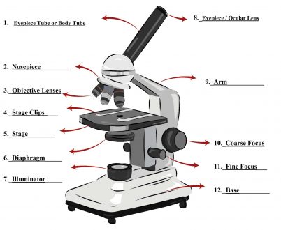
What are the 12 parts of the microscope?
1. Eyepiece or Ocular Lense
Eyepiece lens magnifies the image of the specimen. This part is also known as ocular. Most school microscopes have an eyepiece with 10X magnification.
2. Eyepiece Tube or Body Tube
The tube hold the eyepiece.
3. Nosepiece
Nosepiece holds the objective lenses and is sometimes called a revolving turret. You choose the objective lens by rotating to the specific lens one you want to use.
4. Objective Lenses
Most compound microscopes come with three or four objective lenses that revolve on the nosepiece. The most common objective lenses have power of 4X, 10X and 40X. Combined with the magnification of the eyepiece the resulting magnification is 40X, 100X and 400X magnification. Total magnification is calculated by multiplying the power of the eyepiece by the power of the objective lens. (10X Eyepiece X 40X Objective = 400X Total Magnification) Some more advanced microscopes have an additional objective lens with 100X power. This results in 1,000X magnification. So where do you start? Which objective lens do you need for a particular task? See “How to Use a Compound Microscope” below.
5. Arm
The Arm connects the base to the nosepiece and eyepiece. It is the structural part that is also used to carry the microscope.
6. Stage
The stage is where the specimen is placed. This place is for observation.
7. Stage Clips
Stage clips are the supports that hold the slides in place on the stage.
8. Diaphragm (sometimes called the Iris)
The diaphragm controls the amount of light passing through the slide. It is located below the stage and is usually controlled by a round dial. How to set the diaphragm is determined by the magnification, transparency of the specimen and the degree of contrast you wish to have in your image.
9. Illuminator
Most light microscopes use a low voltage bulb which supplies light through the stage and onto to the specimen. Mirrors are sometimes used instead of a built-in light. If your microscope has a mirror, it provides light reflected from ambient light sources like classroom lights or sunlight if outdoors.
10. Coarse focus
Coarse focus moves the stage to provide general focus on the specimen. When bringing a specimen into focus, the course dial is the first one used.
11. Fine focus
Fine focus moves the stage in smaller increments to provide a clear view of the specimen. When bringing a specimen into focus, the fine focus dial is the second one used.
12. Base
The base is the main support of the microscope. The bottom, where all the other parts of the microscope stand.